Courses
Learning Paths
Step-by-step paths to mastery

5 Levels
5 Courses
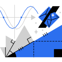
Foundational Math
Master essential skills in Algebra
Browse all 90+ courses


Math
Algebra


Mathematical Thinking


Logic and Deduction


Contest Math

Road to Calculus


Advanced Mathematics


Contributing Authors - Math


Data
Analysis


Probability


Computer Science
Foundational Computer Science


Applied Computer Science


Contributing Authors - CS


Bonus Computer Science Puzzles


Science
Scientific Thinking


Advanced Physics


Contributing Authors - Science


© 2024 Brilliant Worldwide, Inc., Brilliant and the Brilliant Logo are trademarks of Brilliant Worldwide, Inc.